New Insights into Brain Circuits That Regulate Stress and Social Behavior in Mice
UCLA researchers have mapped key brain circuits in mice that control stress responses and social behavior, opening new prospects for targeting neuropsychiatric disorders like depression and PTSD.
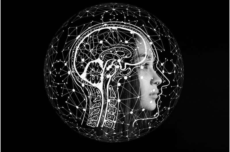
Recent research from UCLA has identified a vital brain hub in mice that plays a crucial role in managing stress responses and social interactions. This groundbreaking study offers a detailed map of neural circuits within the medial prefrontal cortex (mPFC), including the dorsal peduncular area (DP) and infralimbic area (ILA). These regions function as central nodes that integrate sensory input and internal signals, orchestrating emotional and physiological reactions. Understanding how these circuits operate at a cellular level provides valuable insights into the neural basis of emotional stability and stress regulation.
The study, published in the journal Nature, utilized advanced techniques such as genetic labeling, 3D brain imaging, and artificial intelligence-driven circuit mapping to chart these intricate neural networks. The findings reveal that these hubs coordinate complex behaviors and stress responses, and the conserved nature of these circuits suggests they are similar in humans. Lead researcher Dr. Hong Wei Dong emphasized that uncovering this wiring diagram advances our understanding of how the brain governs emotion and social behavior, which could lead to the development of targeted therapies for neuropsychiatric conditions.
Historically, the case of Phineas Gage, a railroad worker whose personality dramatically changed after frontal lobe injury, exemplifies the role of the prefrontal cortex in personality and emotional regulation. Despite this long-standing knowledge, the precise neural pathways involved have remained elusive. This study harnesses innovative genetic and imaging technologies to pinpoint the specific circuits responsible for emotional stability and stress management.
The implications of this research extend to potential clinical applications, offering new avenues for diagnosing and treating disorders such as depression, anxiety, and post-traumatic stress disorder (PTSD). By understanding these core neural networks, scientists aim to develop better, more precise interventions for mental health conditions. Overall, this work marks a significant step toward decoding the brain's complex control systems for behavior and emotional regulation.
Stay Updated with Mia's Feed
Get the latest health & wellness insights delivered straight to your inbox.
Related Articles

The Impact of Physical Activity on Teenagers’ Mental Health: Evidence and Recommendations
Regular physical activity plays a vital role in improving teenagers' mental health by reducing anxiety, depression, and emotional symptoms, while offering long-term psychological benefits.


Understanding Gender Differences in Motivation and Well-Being: Insights from Recent Research
A recent study explores gender differences in motivation and well-being, highlighting the importance of self-efficacy and life meaning across men and women. Discover key insights and practical applications for mental health and personal growth.

The Hidden Dangers of 'What I Eat in a Day' TikTok Videos and Their Impact on Mental Health
TikTok's 'what I eat in a day' videos may seem entertaining but can promote harmful dieting habits and negatively impact mental health. Learn how to navigate this content safely.