Innovative Brain Wave Decoder Enhances Control of Spinal Cord Stimulation

Researchers at Washington University in St. Louis have made significant advancements in restoring communication between the brain and the spinal cord through the development of a novel brain wave decoder. When spinal cord injuries occur, the usual signals from the brain that control movement are interrupted, leading to paralysis. Despite the damage, the brain and the area below the injury retain their normal functions; thus, reconnecting these signals could pave the way for groundbreaking rehabilitation methods.
In their recent study published in the Journal of NeuroEngineering and Rehabilitation, Ismael Seáñez and his team have utilized noninvasive techniques involving electroencephalography (EEG) to capture brain activity. They equipped volunteers with a cap embedded with electrodes to monitor their neural signals while they either physically moved their lower leg or only imagined doing so. This approach allowed the team to gather neural data during both actual movement and mental rehearsals.
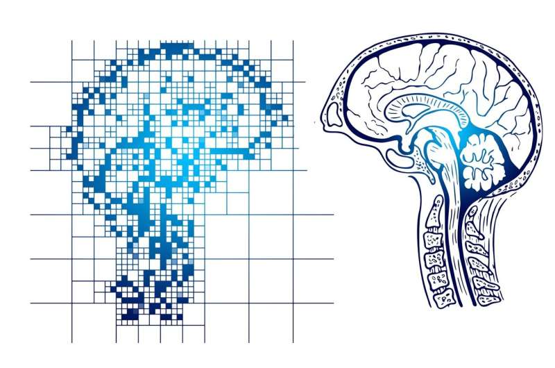
The core of their innovation lies in a specialized decoder algorithm trained to identify patterns of brain activity associated with movement and motor imagery. By providing this neural data to the decoder, it learned to predict when a person was either thinking about moving their leg or actually moving it, even in the absence of physical motion. This capability is crucial because it opens possibilities for using brain signals alone to trigger spinal cord stimulation.
Seáñez explained that the decoder's ability to interpret movement intention from neural activity without actual movement demonstrates its potential for assisting individuals with spinal cord injuries. In future applications, the decoder could be utilized in a brain-spine interface that delivers targeted electrical stimulation transcutaneously—through the skin—to activate spinal circuits and facilitate voluntary movement.
The research indicates that even imagined movement produces neural signatures similar to real movement, which can be harnessed to help restore mobility. The team envisions that a universal decoder trained on aggregated data may simplify clinical application, enabling broader use of this technology in rehabilitation settings.
This promising work represents a significant step toward noninvasive neural interfaces that could revolutionize therapies for spinal cord injury patients by utilizing real-time brain signals to promote functional movements. Further studies aim to refine the decoder for practical use in clinical environments, moving closer to restoring independence for individuals affected by paralysis.
Source: medicalxpress.com
Stay Updated with Mia's Feed
Get the latest health & wellness insights delivered straight to your inbox.
Related Articles
How Sleep Patterns Influence Health, Brain Function, and Lifestyle
New research identifies five distinct sleep profiles linked to brain connectivity, health, and cognition, emphasizing the complexity of sleep and its role in overall well-being.


