Innovation in Brain Imaging: Reversible Transparency Technique in Mice

Stanford researchers have pioneered a reversible, non-invasive technique to make mouse scalps transparent, enabling detailed studies of brain development and neural activity in vivo, opening new horizons in neuroscience research.
Researchers at Stanford University have developed a groundbreaking, reversible method to make mouse scalps transparent, allowing detailed imaging of brain development without invasive procedures. During growth phases in childhood and adolescence, the brain undergoes significant changes, but studying these processes in juvenile mice has been limited due to the challenge of repeatedly observing internal neural pathways. The new technique involves applying a specially formulated solution to the scalp, which temporarily alters its optical properties, turning it transparent to visible light. This enables scientists to observe neural connections, neural activity, and brain structures in live, sedated mice over extended periods.
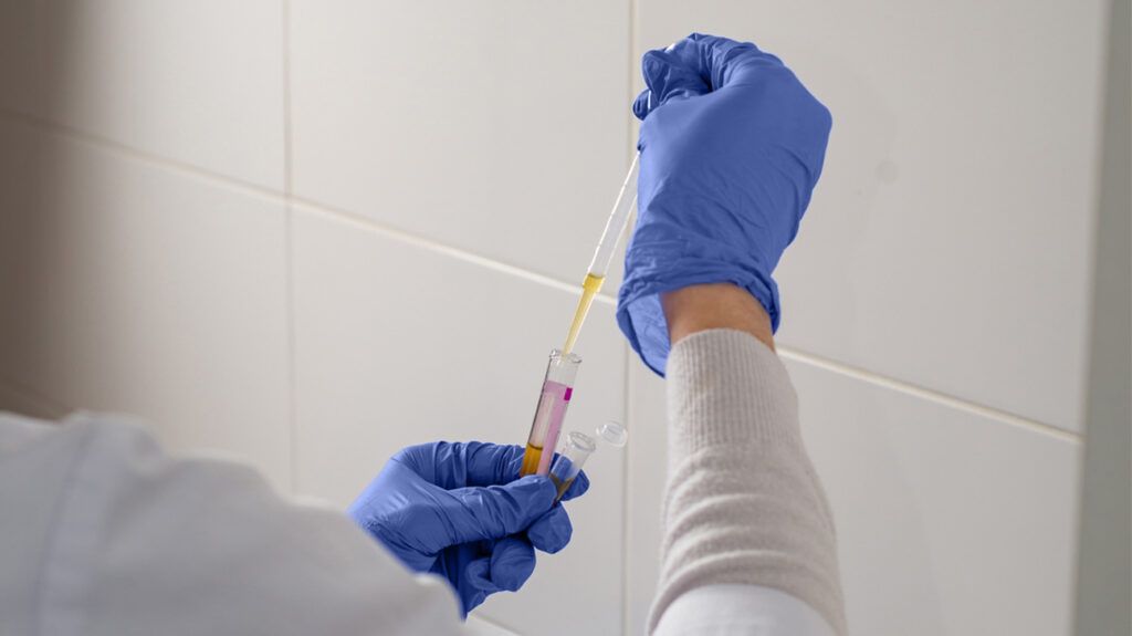
The approach is based on fundamental optical principles. Normally, skin and tissue scatter light because of differences in their refractive indices. The scientific team, including co-author Guosong Hong, achieved transparency by matching the refractive index of the scalp to that of the tissue beneath through the application of ampyrone—a compound that increases the water's refractive index and absorbs ultraviolet light. As a result, light passes through tissues with minimal scattering, revealing internal structures without damaging or permanently altering the tissue.
This breakthrough builds on previous research where the team used similar methods to visualize internal organs through skin transparency. The current process allows imaging across the entire visible spectrum, making it possible to observe fluorescent proteins used for neural activity markers, particularly in young mice with thinner skulls.
Importantly, the transparency effect is temporary—lasting about 20 minutes—and completely reversible, with no apparent harm or irritation to the mice. The researchers are actively seeking more efficient molecules to reduce the required concentration, paving the way for potential translation to human applications—potentially reducing the need for traditional X-ray or CT imaging in medical diagnostics.
By combining physics, chemistry, and neuroscience, this innovative method opens new avenues for longitudinal brain studies, shedding light on how neural circuits form and change during development. This technique holds promise for advancing understanding of neurodevelopmental disorders and improving imaging modalities in biomedical science.
Stay Updated with Mia's Feed
Get the latest health & wellness insights delivered straight to your inbox.
Related Articles

Surge in Whooping Cough Cases in Washington State Amid Declining Vaccination Rates
Washington State is experiencing a sharp rise in whooping cough cases, driven by declining vaccination rates, with severe health risks especially for infants. Learn more about the outbreak and the importance of immunization.

Discovery of Tiny Genetic Segment That Regulates Brain Connectivity and Behavior
A groundbreaking study reveals how a tiny genetic segment, mini-exon B, critically influences brain connectivity, neural balance, and behavior, offering new insights into neurodevelopmental disorders.

Pre-K and Elementary School Students Show Highest Rates of Respiratory Virus Detection
A recent study reveals that pre-kindergarten and elementary students exhibit the highest rates of respiratory virus detection and illness episodes, emphasizing the need for targeted prevention strategies in schools.