Innovative Fluorescent Probe Enables Rapid Visualization of Active Brain Synapses

A groundbreaking fluorescent probe developed by researchers allows rapid visualization of active brain synapses, advancing neuroscience research and understanding of memory formation.
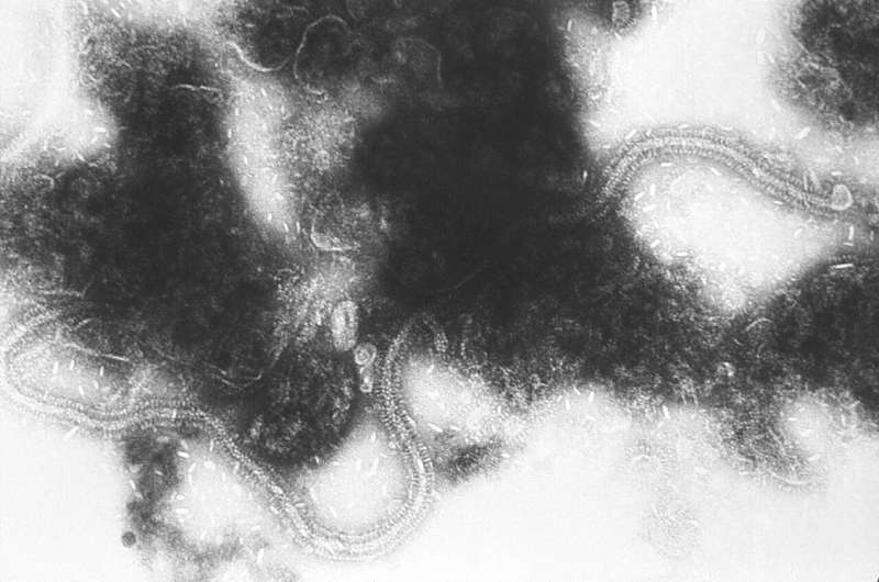
Researchers at Tohoku University and Nagoya University have developed an innovative fluorescent probe that simplifies the process of identifying active brain synapses. The technique involves a straightforward method: just sprinkle the probe onto living brain tissue, and within seconds, active synapses emit light, making them easily visible. This advancement allows scientists to observe how neurons communicate in real time, significantly accelerating neurological research.
Synapses serve as the communication hubs between nerve cells, where neurotransmitters are released to transmit signals. The strength of these synapses influences brain functions such as learning and memory, and this strength can change through a process known as synaptic plasticity. Understanding how synapses adapt during memory formation has long been a goal for neuroscientists.
The new fluorescent probe, called PFQX1(AF488), is specifically designed to bind to AMPA receptors on the surface of neurons. By simply sprinkling the probe on live brain cells, researchers can visually track the localization and movement of AMPA receptors in real time. During experiments involving long-term potentiation (LTP)—a mechanism underlying learning—the team observed that new AMPA receptors are primarily inserted from inside the neuron through exocytosis rather than moving laterally within the membrane.
This discovery was confirmed by blocking exocytosis, which prevented the increase of receptor signals, establishing exocytosis as the main pathway for receptor addition during synaptic strengthening. Notably, this method does not require genetic modifications or complex equipment, making it accessible for widespread use in neuroscience.
The implications of this research are significant. The ability to rapidly visualize and quantify receptor dynamics provides insights into memory formation, brain development, and neurological diseases such as dementia and Alzheimer's. Professor Eriko Nango from Tohoku University states, "Our new probe makes it easier than ever to see which synapses are active, opening new avenues for understanding how the brain learns and adapts."
Overall, this straightforward and efficient technique marks a new chapter in studying the molecular mechanisms of brain function and could pave the way for novel therapeutic approaches to brain disorders.
Stay Updated with Mia's Feed
Get the latest health & wellness insights delivered straight to your inbox.
Related Articles

Innovative Medication Shows Promise in Managing Uncontrolled Hypertension by Targeting Aldosterone Production
A new phase 3 clinical trial highlights lorundrostat, a novel aldosterone synthase inhibitor, as a promising treatment for uncontrolled and resistant hypertension, offering hope for improved management of cardiovascular risk.
Cochrane Review Confirms RSV Vaccines Are Safe and Highly Effective
A new Cochrane review confirms that RSV vaccines are safe and highly effective in protecting infants and older adults from severe respiratory illnesses, showing significant reductions in disease and hospitalization rates.

Surge in Nitrous Oxide-Related Deaths in the US Exceeds 500%, Study Reveals
A study reveals a staggering 500% increase in nitrous oxide-related deaths in the US from 2010 to 2023, raising urgent public health concerns about recreational misuse and its dangers.