Innovative PET Tracer Enables High-Resolution Imaging of Brain Inflammation

A new PET tracer, ¹⁸F-PDE-1905, enables detailed imaging of brain inflammation, offering promise for earlier diagnosis and personalized treatment of neurodegenerative diseases.
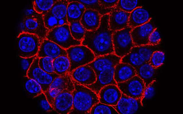
A groundbreaking PET radiotracer has demonstrated the capability to produce detailed images of active brain inflammation in real-time, according to recent research presented at the Society of Nuclear Medicine and Molecular Imaging 2025 Annual Meeting and published in the Journal of Nuclear Medicine. This novel imaging agent, named ¹⁸F-PDE-1905, specifically targets phosphodiesterase 4B (PDE4B), an enzyme involved in inflammatory signaling within microglial immune cells in the central nervous system.
Current PET imaging methods primarily use tracers that focus on TSPO, a marker that is widely expressed across various cell types related to neuroinflammation. In contrast, ¹⁸F-PDE-1905 offers a more specific approach by directly visualizing microglial activation, which is an early indicator of neurodegenerative and psychiatric disorders.
Researchers analyzed genomics data to assess PDE4B expression in diseases such as Parkinson's and multiple sclerosis. They developed mouse models of neuroinflammation and performed dynamic PET scans using ¹⁸F-PDE-1905, alongside the traditional TSPO-specific tracer ¹⁸F-D2-LW223. The results showed higher uptake of the new tracer in diseased brains, indicating increased inflammation. Notably, ¹⁸F-PDE-1905 provided superior image clarity and broader brain distribution, highlighting its potential for precise neuroinflammation imaging.
Bioinformatics analysis confirmed elevated PDE4B levels in both human patients and mouse models of neuroinflammatory diseases. This increased enzyme activity was correlated with higher tracer uptake in imaging studies. By targeting PDE4B directly, ¹⁸F-PDE-1905 can visualize inflammatory processes more accurately and earlier than existing methods.
This advancement in PET imaging technology could lead to earlier diagnosis, better monitoring of disease progression, and tailored therapies for conditions like Alzheimer's, Parkinson's, ALS, and multiple sclerosis. The approach signifies a shift towards more targeted, personalized treatment strategies in neurology, emphasizing the importance of precise neuroinflammation assessment for improved patient outcomes.
Stay Updated with Mia's Feed
Get the latest health & wellness insights delivered straight to your inbox.
Related Articles

Disparities in Flu Vaccination Rates Highlight Racial Gaps
A study shows persistent racial disparities in flu vaccination rates, with Asian patients consistently having higher uptake compared to Black patients across all age groups and flu seasons.
Innovative Vaccines Show Promise in Combating Pancreatic Cancer in Preclinical Studies
New vaccine therapies targeting pancreatic cancer show promising results in preclinical studies, offering hope for more effective treatments or prevention methods against this deadly disease.

Artificial Intelligence Improves Diagnostic Precision for High-Risk Thyroid Nodules
A new deep learning model combining ultrasound techniques significantly improves the accuracy of diagnosing high-risk thyroid nodules, aiding clinicians in early and precise detection of thyroid cancer.

Breakthrough HIV Neutralizing Antibody Shows Promise in Laboratory Tests
A newly discovered antibody shows remarkable potential in neutralizing over 98% of HIV strains, promising advances in HIV prevention and therapy efforts.